Aluminium »
PDB 5c2k-6i03 »
5gza »
Aluminium in PDB 5gza: Protein O-Mannose Kinase
Enzymatic activity of Protein O-Mannose Kinase
All present enzymatic activity of Protein O-Mannose Kinase:
2.7.1.183;
2.7.1.183;
Protein crystallography data
The structure of Protein O-Mannose Kinase, PDB code: 5gza
was solved by
J.Xiao,
with X-Ray Crystallography technique. A brief refinement statistics is given in the table below:
| Resolution Low / High (Å) | 35.28 / 2.00 |
| Space group | P 32 |
| Cell size a, b, c (Å), α, β, γ (°) | 70.550, 70.550, 66.935, 90.00, 90.00, 120.00 |
| R / Rfree (%) | 19.7 / 21.8 |
Other elements in 5gza:
The structure of Protein O-Mannose Kinase also contains other interesting chemical elements:
| Fluorine | (F) | 3 atoms |
| Magnesium | (Mg) | 2 atoms |
Aluminium Binding Sites:
The binding sites of Aluminium atom in the Protein O-Mannose Kinase
(pdb code 5gza). This binding sites where shown within
5.0 Angstroms radius around Aluminium atom.
In total only one binding site of Aluminium was determined in the Protein O-Mannose Kinase, PDB code: 5gza:
In total only one binding site of Aluminium was determined in the Protein O-Mannose Kinase, PDB code: 5gza:
Aluminium binding site 1 out of 1 in 5gza
Go back to
Aluminium binding site 1 out
of 1 in the Protein O-Mannose Kinase

Mono view
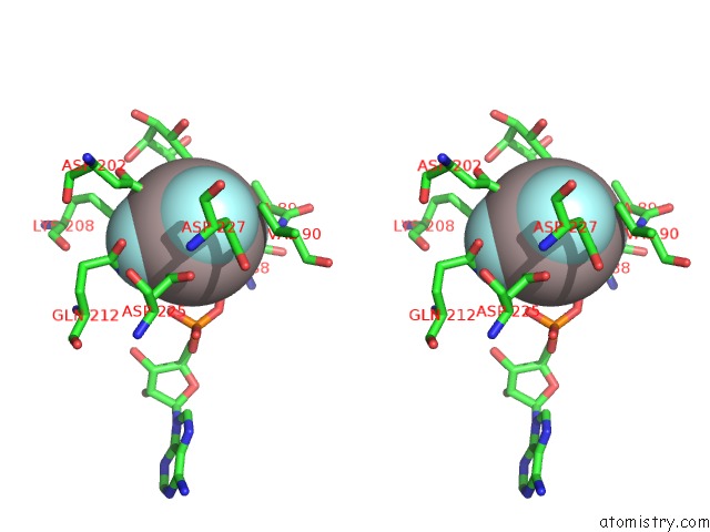
Stereo pair view
Mono view
Stereo pair view
A full contact list of Aluminium with other atoms in the Al binding
site number 1 of Protein O-Mannose Kinase within 5.0Å range:
|
Reference:
Q.Zhu,
D.Venzke,
A.S.Walimbe,
M.E.Anderson,
Q.Fu,
L.N.Kinch,
W.Wang,
X.Chen,
N.V.Grishin,
N.Huang,
L.Yu,
J.E.Dixon,
K.P.Campbell,
J.Xiao.
Structure of Protein O-Mannose Kinase Reveals A Unique Active Site Architecture Elife V. 5 2016.
ISSN: ESSN 2050-084X
PubMed: 27879205
DOI: 10.7554/ELIFE.22238
Page generated: Wed Jul 10 09:49:57 2024
ISSN: ESSN 2050-084X
PubMed: 27879205
DOI: 10.7554/ELIFE.22238
Last articles
Zn in 9MJ5Zn in 9HNW
Zn in 9G0L
Zn in 9FNE
Zn in 9DZN
Zn in 9E0I
Zn in 9D32
Zn in 9DAK
Zn in 8ZXC
Zn in 8ZUF